
Share this
October 2, 2020
Nanotechnology is increasingly used in life sciences research, with applications in diagnostics, imaging, and therapeutics, just to name a few. Nanoparticulates are difficult to study due to their size and the limitations of conventional optical imaging technologies. For researchers to fully gauge the efficacy and toxicity of nanoparticles, it’s imperative to study the design, structures, and behaviors of nanoparticles in vivo and in 3D.
At the Institut Lumiere in France, the researchers recently shared their proof-of-concept study that will help demonstrate the use of LIBS in the development of 3D label-free nanoparticle imaging at the entire-organ scale. Quantum Composers instruments were used in this study and they are honored to be a part of this groundbreaking research. These findings have the potential to help advance gene therapies, drug delivery, tumor targeting, and much more. Read on to explore this amazing new LIBS application.
LIBS in Biological Imaging
LIBS may not be the most sensitive or high-resolution option, but LIBS offers the advantage of a fast operating speed and an all-optical tabletop instrument that is compatible with standard optical microscopy. The scanning speed can be up to 100 times faster than other techniques, allowing 3D investigations to be conducted on large biological samples within reasonable time periods. LIBS, combined with a volume reconstruction of a sliced organ and in-depth analysis, demonstrated for the first time that LIBS imaging can be used for the 3D imaging of biological organs.
Attenuator Module Stabilizes Laser Energy
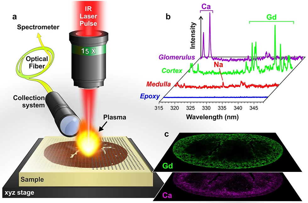
The LIBS experiment at the Lumiere Institute used Nd:YAG laser pulses of 1064 nm, focused onto a sample by a 15x magnification objective using a pulse duration of 5 ns and a repetition rate of 10 Hz. A beam shutter was used to control the delivery of the laser pulse to the sample such that only one plasma plume was produced for each position of the sample. To perform the mapping experiments at the greatest possible speed, the movement of the sample was synchronized with the opening of the beam shutter, and the spatial resolution was set by adjusting the speed of the motorized three-axis translation stage. Quantum Composers’ computer-controlled attenuator (model ATT1064) provided linearized control of the laser’s energy. Monitoring the laser output with a power meter a servo control loop was used with the attenuator to control the laser energy, which improved the long-term stability of the laser output. The attenuator’s internal calibration and linearization function simplified setup and calibration.
At Quantum Composers, we are proud to have our instrument chosen for use in this study, which demonstrates that LIBS is suitable for label-free, nanoparticle 3D imaging of biological tissue within entire organs. You’re invited to view the attenuator instrument and the entire study at the links below. Our team has a rich history of working with engineers and scientists in academia around the world, synchronizing hardware for demanding and complex experiments.
Gimenez, Y. et al. 3D Imaging of Nanoparticle Distribution in Biological Tissue by
Laser-Induced Breakdown Spectroscopy. Sci. Rep. 6, 29936; doi: 10.1038/srep29936 (2016).